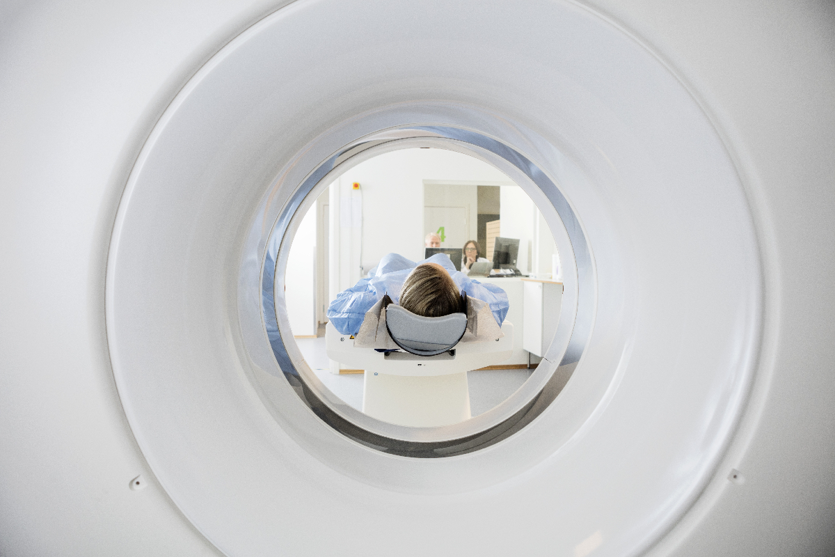
High-resolution computed tomography is the most common imaging, apart from chest X-ray, used to help assess severe asthma. There are numerous published reviews available:
The predominant use of HRCT is exclusion of alternate or coexistent diagnoses (e.g. bronchiectasis, emphysema, mucous plugging, fibrosis, paralysed hemidiaphragm, idiopathic interstitial pneumonia including eosinophilic pneumonia, ABPA).
Minor abnormalities assessed by clinical radiological interpretation, are common. These include (Richards et al. 2016)
- Bronchial dilatation
- Bronchial wall thickening
- Mucous plugging
- Ground glass opacities
- Mosaic perfusion
- Expiratory gas trapping
Advanced image analysis techniques are currently being developed which may provide useful functional information from HRCT in asthma (e.g. PRM – parametric response mapping)
Other imaging modalities are in the research area only:
- Examples: Optical coherence tomography, hyperpolarised He3 MRI, Single Photon Emission Computed Tomography, Positron Emission Tomography, oxygen enhance MRI (e.g. SVI – specific ventilation imaging), endobronchial ultrasound.

